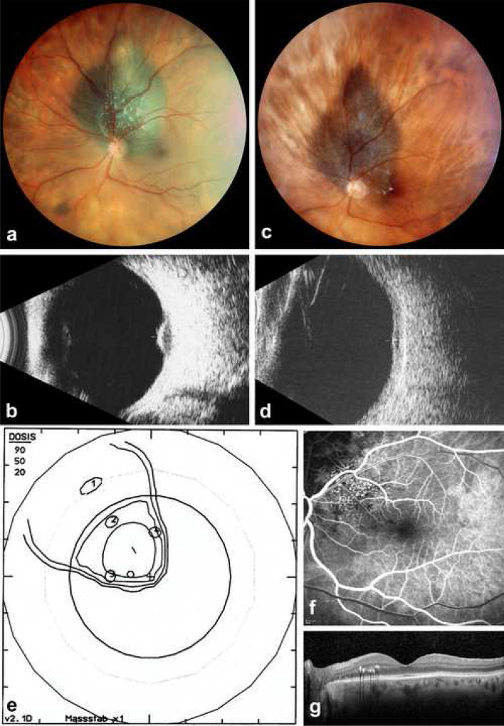
Fig. 1.
Parapapillary choroidal melanoma (left eye) maintaining useful vision following proton therapy. a Panoramic fundus photo (Panoret camera) in a 71-year-old female at initial presentation with loss of VA to 0.6 decimal related to a secondary macular detachment. b On B-scan ultrasonography (10 MHz), thickness is 3.4 mm. c Eleven years after radiation therapy, the tumor borders are under control on panoramic fundus photography, with some lipid exudates close to the macular border. d On B-scan ultrasonography (10 MHz), the atrophic scar has a residual thickness of 1.3 mm. e Proton therapy irradiation plan—fundus view—illustrating the tumor base, localized by four tantalum clips and surrounded by the 90, 50 and 20% isodose borders. f Fluorescein angiography (early venous phase) of the macula, on which radiation-induced extrafoveal telangiectasia can be identified, explaining the lipid exudates. g On B-scan OCT, the fovea appears normal, with some extrafoveal lipid exudates. VA is 1.0 decimal

