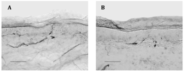
Figure 1.

Representative skin biopsies from 10 cm above the lateral malleolus, bright-field, 40 x. Scale bar =100μm; arrows demonstrate epidermal nerve fibers
A. Healthy control: Biopsy from 17.9-year-old Caucasian female with epidermal neurite density (END) of 500/mm2 skin surface area; at the 64.3 percentile of the age, sex, and race-adjusted normal distribution.
B. Juvenile fibromyalgia patient: Biopsy from a 17.8-year-old Caucasian patient with END (242/mm2 skin surface area) at the 4.7 centile of predicted.
