Figure 5.

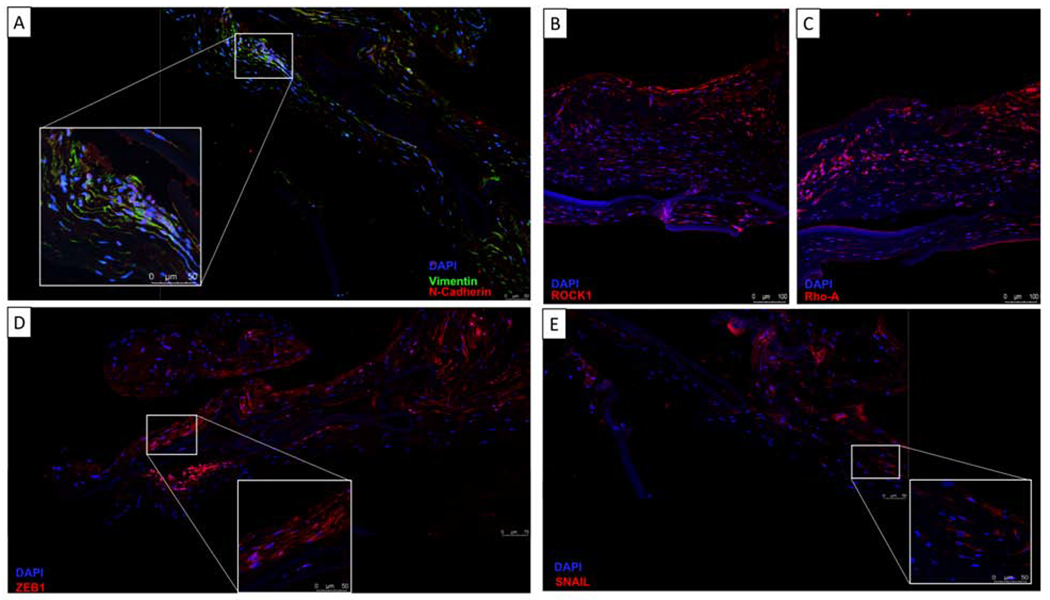
Retrocorneal membrane immunofluorescence on paraffin-embedded sections. Membranes demonstrate positivity for Vimentin (A), N-Cadherin (A), Rock1 (B), Rho-A (C), Zeb1(D) and Snail (E). All nuclei were counterstained with DAPI (blue).

Retrocorneal membrane immunofluorescence on paraffin-embedded sections. Membranes demonstrate positivity for Vimentin (A), N-Cadherin (A), Rock1 (B), Rho-A (C), Zeb1(D) and Snail (E). All nuclei were counterstained with DAPI (blue).