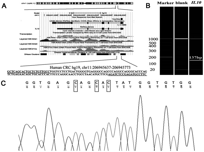
Figure 1.
Target sequence on IL10 promoter region. (A) Genomic position and functional annotation of the amplified fragment. The primers for the quantitative methylation-specific PCR were underlined, and five CpG sites were identified (in grey). (B) Capillary electrophoresis of the amplified fragment (137 bp). (C) Sanger sequencing results. Top row of the sequence represents the original sequence of the fragment. Second row of the sequence represents the converted sequences. C nucleobase with corresponding converted T were in black boxes. A, adenine; C, cytosine; F, forward; G, guanine; IL10, interleukin 10; R, reverse; T, thymine.

