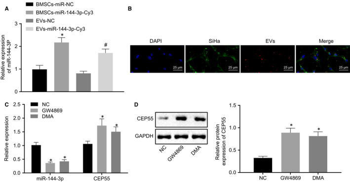
FIGURE 6.

miR‐144‐3p can be transferred into cervical cancer cells by EVs derived from hBMSCs. A, Expression of miR‐144‐3p was determined by RT‐qPCR in cells, relative to U6. B, miR‐144‐3p‐Cy3 in hBMSCs and cervical cancer cells under the fluorescence microscope (scale bar = 20 µm). C, Expressions of miR‐144‐3p and CEP55 were determined by RT‐qPCR in cells, relative to U6 and GAPDH, respectively. D, Representative Western blots of CEP55 protein and its quantitation in cells, normalized to GAPDH. Data comparison was analysed by independent sample t test between two groups and by one‐way ANOVA among multiple groups, followed by Tukey's test. *P < 0.05 versus hBMSCs treated with miR‐NC or NC. # P < 0.05 versus EVs treated with NC. Data are shown as the mean ± standard deviation of three technical replicates
