
FIGURE 5.
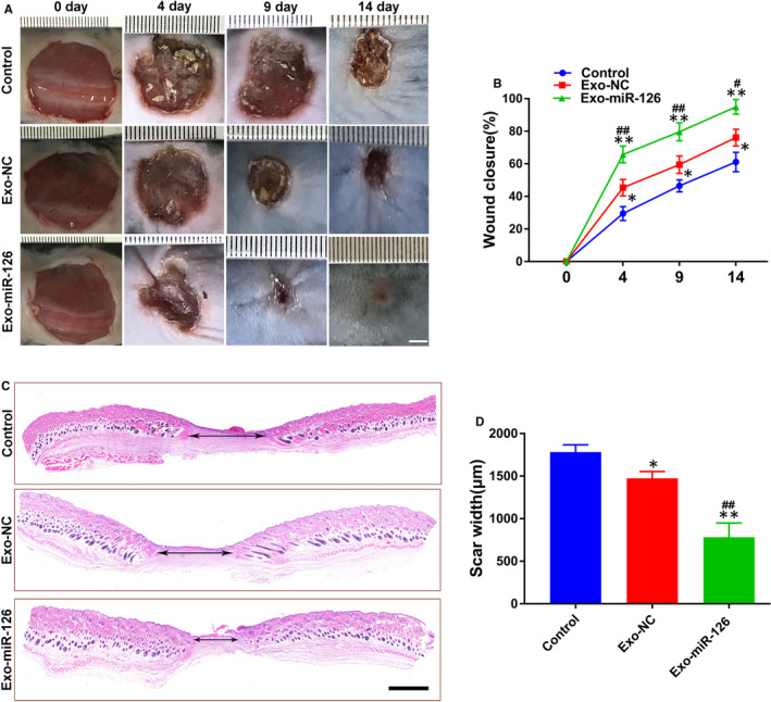
Exo‐miR‐126 accelerated cutaneous wound healing in mice. (A) Gross view of wounds in mice receiving different treatments at days 4, 9 and 14 post‐wounding (scale bar: 2 mm). (B) The rate of wound closure in wounds receiving different treatments at the indicated times (n = 8). (C) H&E staining of wound sections from mice receiving different treatments at 14 days after operation. The double‐headed black arrows indicate the edges of the scars (scale bar: 500 μm). (D) Quantitative analysis of scar widths in (C) (n = 8). *P < 0.05, **P < 0.01 vs. Control group, # P < 0.05, ## P < 0.01 vs. Exo‐NC group. Exo‐NC, exosomes derived from BMMSCs transfected with NC‐mimic; Exo‐miR‐126, exosomes derived from microRNA‐126 overexpressing bone marrow mesenchymal stem cells