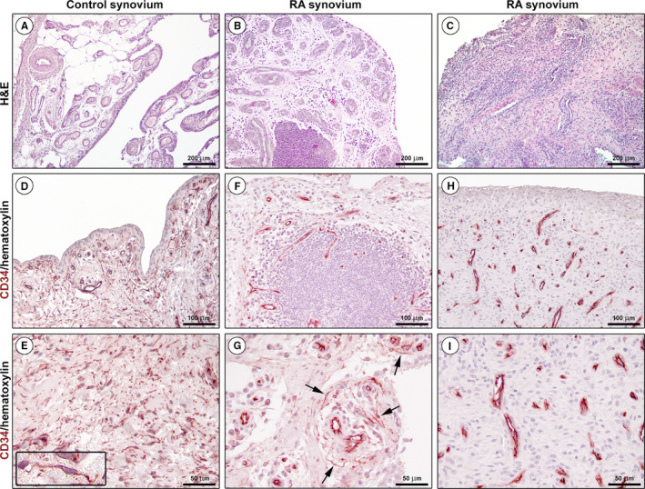
FIGURE 1.

Representative light microscopy photomicrographs of synovial tissue sections from healthy controls and patients with rheumatoid arthritis (RA). (A‐C) Haematoxylin and eosin (H&E) staining. Healthy synovium consists of a thin intimal lining layer and a sublining layer of vascularized loose connective tissue with only very few leucocytes (A). Hyperplastic RA synovium displays either diffuse immune cell infiltrates or ectopic lymphoid structures in the sublining layer (B, C). (D‐I) CD34 immunohistochemistry with haematoxylin counterstain. An intricate network of telocytes (TCs)/CD34+ stromal cells is present in the whole sublining layer of healthy control synovium (D, E). At higher magnification, TCs/CD34+ stromal cells appear as spindle‐shaped cells with a small nucleated body and long cytoplasmic processes with an irregular calibre (inset in E). CD34 immunoreactivity is observed also in endothelial cells of blood microvessels. Note the presence of a few perivascular TCs/CD34+ stromal cells in the sublining layer of a RA synovial sample (F, arrows in G). Note the complete absence of TCs/CD34+ stromal cells in another RA synovial sample; all CD34+ tissue structures are identifiable as blood microvessels (H, I). Scale bar: 200 μm (A‐C), 100 μm (D, F, H), 50 μm (E, G, I)
