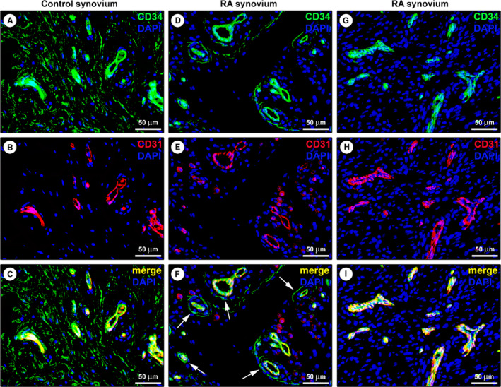
FIGURE 2.

Representative fluorescence microscopy photomicrographs of synovial tissue sections from healthy controls and patients with rheumatoid arthritis (RA). (A‐I) Double immunofluorescence labelling for CD34 (green) and CD31 (red) with DAPI (blue) counterstain for nuclei. In healthy control synovium, telocytes (TCs)/CD34+ stromal cells lacking CD31 immunoreactivity form an extensive network in the sublining layer; endothelial cells of blood microvessels are CD31+/CD34+ (A‐C). Note the presence of a few CD31−/CD34+ TCs around CD31+/CD34+ blood microvessels in the sublining layer of a RA synovial sample (D‐F, arrows in F). In another RA synovial sample, the sublining layer displays numerous CD31+/CD34+ blood microvessels, but CD31−/CD34+ TCs are undetectable (G‐I). Scale bar: 50 μm (A‐I)
