Figure 6.

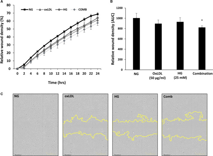
High glucose (HG), OxLDL and combination of both decreases endothelial cell migration in scratch wound assay. HMEC‐1 cells were incubated with either HG or NG for 4 d and with or without OxLDL (50 µg/mL) for additional 24 h. Then, cells were wounded by automated scratcher at 100% confluence. Medium was switched from 10% FCS to starvation mode (2% FCS) 2‐3 h prior to scratch performance and throughout scratch evaluation. Phase‐contrast images of wounded areas were taken each hour over a period of 24 h using IncuCyte ZOOM® system. A, Relative wound density of the wound of each group over 24 h. B, Area under the curve of the wound density. C, Representative images of the wound of each cell type and each treatment at 24 h. *P < .05
