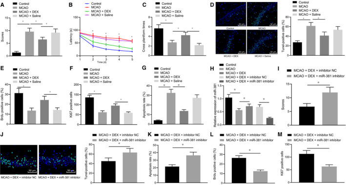
Figure 1.

Dex enhances the miR‐381 expression to alleviate cerebral ischaemic injury in the MCAO rat. A, The mNSS score of rats after MCAO model establishment and Dex injection. B‐C, Latency for searching for the hidden platform and number of crossings the platform of rats after MCAO model establishment and Dex injection. D, TUNEL staining on brain tissues of rats after MCAO model establishment and Dex injection. E, BrdU‐positive cell rate was detected by BrdU. F, Ki‐67 staining to detect the number of Ki‐67‐positive expression. G, Flow cytometry analysis of cell apoptosis in brain tissues of rats after MCAO model establishment and Dex injection. H, miR‐381 expression of rats after MCAO model establishment, Dex injection and miR‐381 inhibitor treatment detected by RT‐qPCR (normalized to U6). I, The mNSS score of rats. J, TUNEL staining results of brain tissues of rats (scale bar = 100 μm). K, Apoptotic cells in brain tissues of rats measured by TUNEL staining. L, Cell apoptosis in brain tissues of rats determined by flow cytometry. N, BrdU‐positive cell rate was detected by BrdU. M, Ki‐67 staining to detect the number of Ki‐67‐positive expression. *P < .05. The above measurement data were expressed as the mean ± SD. The data of multiple groups were compared by one‐way ANOVA and Tukey's post hoc test. Data comparisons at different time‐points were performed by repeated‐measures ANOVA and Bonferroni's post hoc test. Non‐parametric test (Mann‐Whitney U test) was used to compare the neurological scores. n = 12
