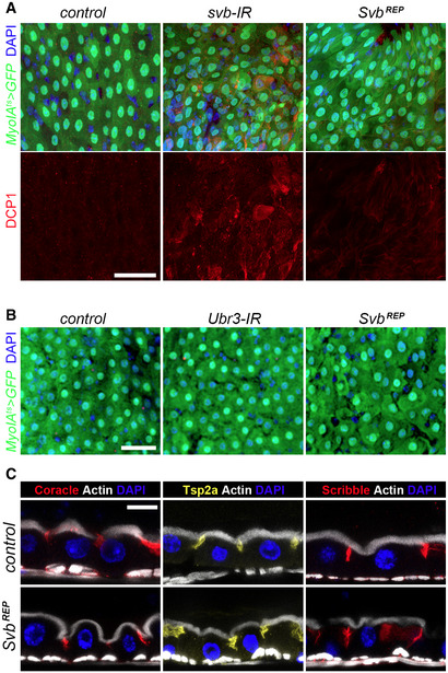
Figure EV5. Svb acts as a transcriptional repressor in differentiated enterocytes.

- Myo1Ats midguts expressing GFP alone (control), or expressing svb‐RNAi and SvbREP. Samples were stained for GFP (green) and DAPI (blue). Lower panel shows staining for cleaved DCP1 (red). Scale bar is 20 µm.
- Myo1Ats midguts expressing GFP alone (control), or expressing Ubr3‐RNAi and SvbREP. Samples were stained for GFP (green) and DAPI (blue). Scale bar is 20 µm.
- Cross sections of control MyoIAts midguts (expressing GFP and mCherry‐RNAi, top row), or SvbREP (bottom row). Samples were stained for F‐actin (white), DAPI (blue), and Coracle (red), Tsp2a (yellow) or Scribble (red). Scale bar is 5 µm.
