Figure 2.

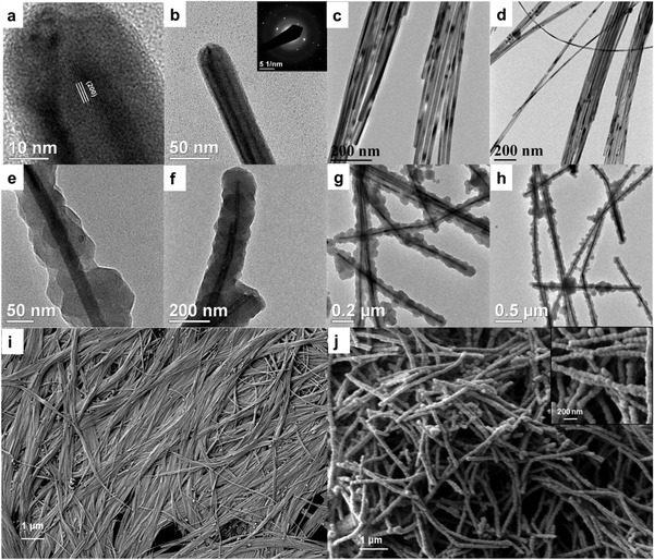
TEM images of a–d) Cu NWs, e–h) Cu@ZIF‐8 NWs. The inset in (b) shows a representative SAED pattern from a Cu NW. SEM images of i) Cu NWs, j) Cu@ZIF‐8 NWs.

TEM images of a–d) Cu NWs, e–h) Cu@ZIF‐8 NWs. The inset in (b) shows a representative SAED pattern from a Cu NW. SEM images of i) Cu NWs, j) Cu@ZIF‐8 NWs.