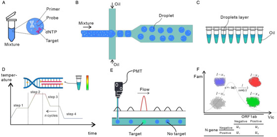
FIGURE 1.

Droplet digital PCR workflow. (A) A mixture containing dNTPs, primers, and probes is prepared for the amplification reaction. (B) Water‐in‐oil droplets are generated using a microfluidic flow‐focusing system. (C) The generated droplets and oil are collected in PCR tubes. (D) The PCR tubes are placed in a thermal cycler; PCR amplification occurs in each droplet. (E) After amplification, the droplets are checked by a photoelectric detection system composed of lasers and photomultiplier tubes. The fluorescence of droplets containing target molecules is strong enough to be distinguished from the background fluorescence. (F) Finally, the fraction of positive droplets is fitted to the Poisson distribution to determine the absolute copy number of target molecules in the original reaction mixture
