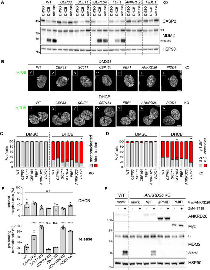
Figure 3. PIDD1 localization to DAs is necessary for PIDDosome activation.

- RPE1 cells of the indicated genotypes were treated either with DMSO or with DHCB for 24 h. A fraction of DHCB‐treated cells were released into fresh medium for other 24 h (release). Samples were subjected to immunoblotting; n = 3 independent experiments.
- Fluorescence micrographs of RPE1 cells of the indicated genotypes, either treated for 24 h with DHCB or with vehicle alone (DMSO). Blow‐up without Hoechst 33342 is magnified 2.5×. Scale bar: 5 μm.
- Immunofluorescence micrographs of RPE1 as in (B) were used to visually assess the percentage of cells presenting one or two nuclei. N = 3, ≥ 50 cells from each independent experiment. Mean values ± s.e.m. are reported. The increase in the number of binucleated cells between the wild‐type sample and all the other genotypes was assessed (ANOVA test; *P < 0.05).
- Immunofluorescence micrographs of RPE1 as in (B) were used to visual score the number of centrosomes per cell by counting the number of γ‐tubulin‐positive centrioles. N = 3, ≥ 50 cells from each independent experiment. Mean values ± s.e.m. are reported. The increase in the number of cells with > 2 centrosomes between the wild‐type sample and all the other genotypes was assessed (ANOVA test; **P < 0.01).
- Quantitative assessment of the fraction of RPE1 cells undergoing cytokinesis failure upon DHCB treatment, inferred on the basis of the increase of ploidies ≥ 4C (upper panel) and of the fraction of the abovementioned cells undergoing genome reduplication upon release after DHCB treatment (lower panel). Individual values of biological replicates, their mean and standard deviations are reported. ANOVA test, comparing each sample to the wild type (****P < 0.0001; n.s. = non‐significant).
- RPE1 cells were either left untransduced (mock) or transduced with lentiviral vectors expressing the indicated Myc‐ANKRD26 constructs. Subsequently, cells were treated either with DMSO or ZM447439 for 24 h and subjected to immunoblotting; n = 3 independent experiments.
Source data are available online for this figure.
