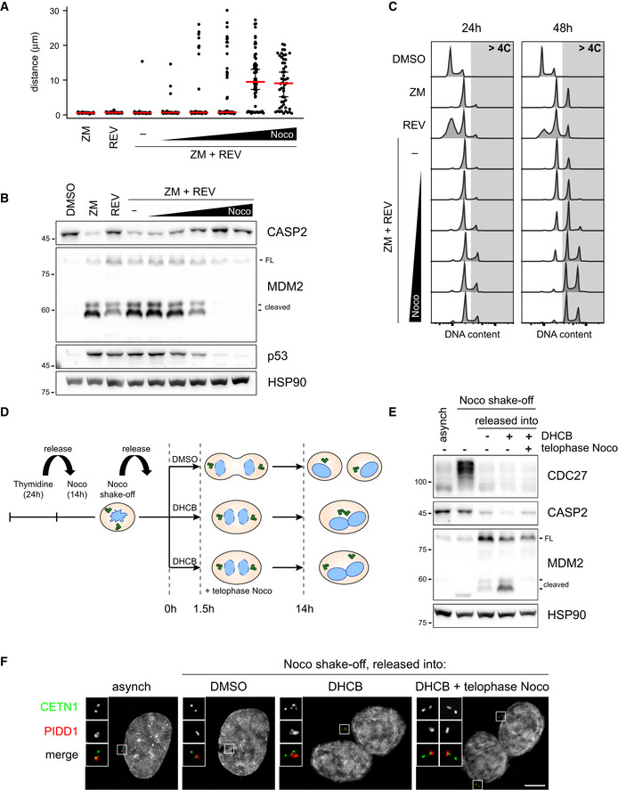
Figure 7. Centrosome clustering is necessary for PIDDosome activation.

- Dot plot showing the distance between parent centrioles pairs in A549 cells following the indicated treatments (ZM = ZM447439; REV = reversine). Nocodazole concentrations are 0.03, 0.1, 0.33, 1, 3.3 µM. Median (red) and 95% confidence interval thereof (black) are shown. N > 50 cells were analysed.
- Immunoblot of A549 cells subjected to the indicated treatments for 24 h as in (A). N = 3 independent experiments.
- DNA content analysis of A549 cells subjected to the indicated treatments as in (A) either for 24 h (left panels) or 48 h (right panels). N = 2 independent experiments.
- Schematic of the experimental conditions utilized to synchronize RPE1 cells and to specifically interfere with centrosome clustering after telophase.
- Immunoblots of RPE1 cells synchronized as in (D). N = 3 independent experiments.
- Representative fluorescence micrographs of RPE1 cells synchronized as in (D). Centrosomal antigens were stained with the indicated antibodies. Blow‐ups without Hoechst 33342 are magnified 2×. Scale bar: 5 μm.
Source data are available online for this figure.
