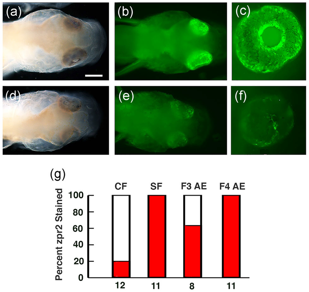
FIGURE 4.

Retinal photoreceptor layer in the albino eyed (AE) strain determined by staining with zpr2 antibody at 3 days post fertilization. (a-c) The majority of F3 AE larvae stain positively for zpr2 antigen. (d-f) A minority of F3 AE larvae lack appreciable zpr2 staining. (a, d) Bright field images. (b, c, e, f) Fluorescence images. Scale bar in (b)=300μm; magnifications are the same in (a-f). (g) Bar graphs showing the percentage (dark bars) of cavefish (CF), surface fish (SF), F3 AE, and F4 AE with eyes showing zpr2 staining. Number of samples assayed is shown at the bottom of each bar
