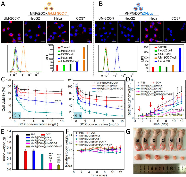
Figure 13.
(A,B) Increased internalization of DOX-loaded magnetic iron oxide NPs (MNPs) coated with membrane fragments of UM-SCC-7 (A) and HeLa (B) cells in homotypic cancer cells compared to other cell lines. (C) Viability of UM-SCC-7 cells upon 3 and 6 h exposure to DOX-loaded MNPs coated with membranes obtained from different cell lines (UM-SCC-7, COS7, and HeLa). (D,F) Relative tumor volumes (D) and body weights (F) upon treatment of UM-SCC-7 tumor-bearing mice with variations of MNPs with or without targeting by application of an external magnetic field (MF) on days indicated by the red arrows. (E) Average tumor weight measured on day 12 post treatment. (G) Photograph of UM-SCC-7 tumor-bearing mice and tumors on day 12 post treatment for each of the different groups (a = PBS, b = DOX, c = @HeLa, d = @COS7, e = @UM-SCC-7, f = @UM-SCC-7 + MF). Reproduced with permission from ref (342). Copyright 2016 American Chemical Society.

