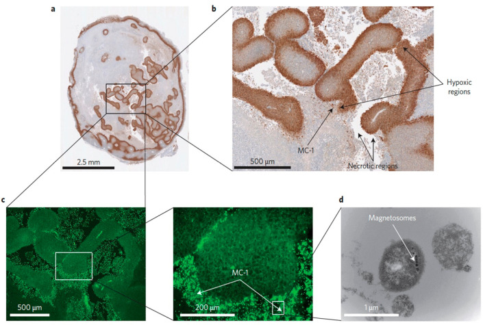
Figure 14.
(a,b) Peritumorally injected MC-1 cells localize in hypoxic regions (brown islands) of mouse xenografts. (c) Fluorescent images of MC-1 bacteria stained with FITC-conjugated secondary antibodies in adjacent sections of the same xenografts. (d) TEM images of MC-1 bacteria highlighting the presence of magnetosomes. Reproduced with permission from ref (365). Copyright 2016 Nature Publishing Group.

