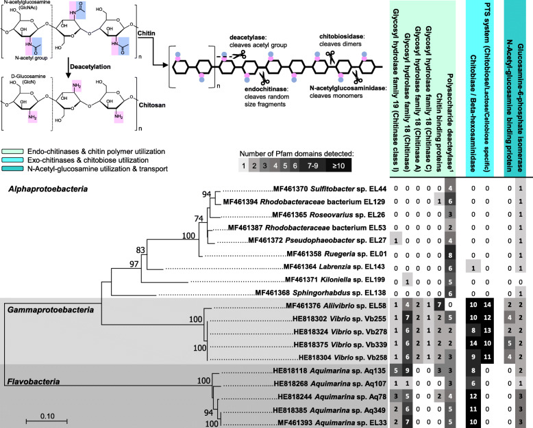
Fig. 1.
Annotation of chitin and chitin-derivative degradation and utilization genes in cultivated bacterial symbionts of sponges and octocorals. Nineteen genomes available from the panel of 41 strains examined in this study are portrayed, spanning ten formally described and two potentially novel genera across three bacterial classes. The phylogenetic tree on the bottom left is based on a maximum likelihood analysis (Generalized Time Reversible model) of the 16S rRNA gene (16S rRNA gene accession numbers are given next to each strain name). Numbers at nodes represent values based on 300 bootstrap replicates. The table on the right shows Pfam-based annotations for predicted protein domains involved in hydrolysis (endo-chitinases of GH families 18 and 19—EC 3.2.1.14, chitin-binding proteins) and deacetylation (polysaccharide deacetylases) of the of the large chitin polymer (light green panel), hydrolysis (exo-chitinases, EC 3.2.1.52) of chitin non-reducing ends and chitobiose transport (light blue panel), and N-acetylglucosamine binding and utilization (blue panel); the latter function represented by the presence of glucosamine 6-phosphate isomerases (EC 3.5.99.6). These functional categories have been as well used to estimate the relative abundance of CDSs involved in the breakdown and utilization of chitin and chitin derivatives across the sponge and octocoral metagenome datasets (Fig. 3). Values in each cell of the table correspond to the number of Pfam domains detected for each functional category in each genome, whereby higher numbers are highlighted in dark-gray shading. The upper left panel shows the molecular structure of chitin and chitosan. It highlights the N-acetyl group characteristic of the chitin polymer, which is cleaved by deacetylases in the process of chitosan formation, and the cleaving sites of endo- and exo-chitinases along the chitin chain. Superscript “1” corresponds to InterPro database entry IPR002509 (see also Fig. 3) which describes the metal-dependent deacetylation of O- and N-acetylated polysaccharides such as chitin, peptidoglycan, and acetylxylan

