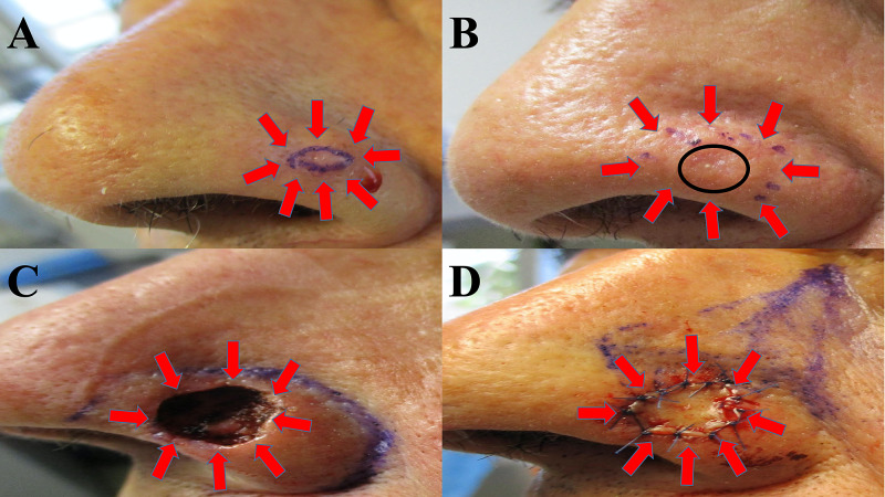
Figure 1. Clinical presentation of calcifying basal cell carcinoma and removal with Mohs microscopically controlled surgery.
An 80-year-old man presented with a nodule on his left nasal ala (A); the tumor is surrounded by red arrows, and the biopsy site is the skin within the purple oval. The lesion site was injected with epinephrine-containing anesthetic before the shave biopsy, accounting for whitening of the surrounding tissue. The biopsy site (B) was healed at four weeks follow-up (black circle), and Mohs surgery was scheduled to excise the nodular basal cell carcinoma with calcinosis cutis (surrounded by red arrows). After three stages of Mohs micrographic surgery, clear tumor margins were obtained; the resulting skin defect on the left nasal ala is surrounded by red arrows (C). A skin-advancing flap was initially planned (purple lines demarcating the planned incisions) to close the nasal defect (D); however, a full thickness skin graft (surrounded by red arrows) from posterior auricular skin was used instead.

