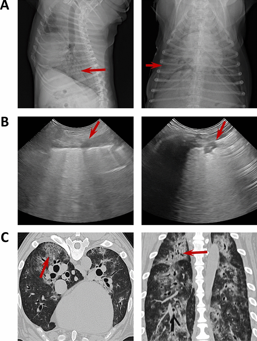
Figure 1.

Imaging with chest radiograph, sonographic images and CT of three different dogs. A Thoracic radiograph made in right lateral (left) and dorsoventral (right) showing a generalized severe interstitial opacity accentuated in the caudodorsal (arrows). B Sonographic images of two patients with severe dyspnea showing a diffused B line (left; arrow) and consolidation focal lesions (right; arrow). C Transverse (left) chest CT images showing bilateral focal peripheral ground-glass opacities with intralobular and interlobular smooth septal thickening (arrow); sagittal (right) chest CT images showing diffuse opacities with consolidation and bronchial wall thickening (arrow).
