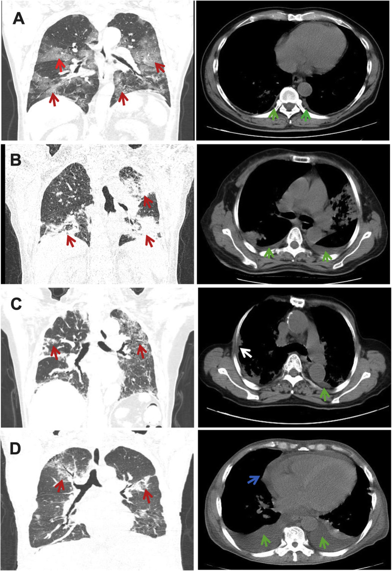
Fig. 2.

Imaging findings of COVID-19 patients with pleural effusion: a: Multifocal ground-glass opacities (red arrow on the coronal image) and pleural effusion (green arrow on the axial image). b: Multiple patchy consolidation in the upper left lobe (red arrow on the coronal image) and the lower two lobes with bilateral pleural effusion (green arrow on the axial image). c: Bilateral ground-glass opacities (red arrow on the coronal image) and pleural effusion (green arrow on the axial image) with pleural thickening (white arrow on the axial image). d: Bilateral ground-glass opacities (red arrow on the coronal image) with pleural (green arrow on the axial image) and pericardial effusion (blue arrow on the axial image)
