
Figure 3.
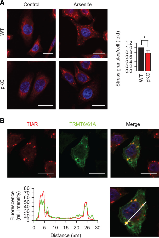
TRMT6/61A is involved in stressed-induced granulation. (A) Immunofluorescence staining of TIAR (red) to detect SG formation upon arsenite treatment for 30 min in serum-free medium. DAPI staining (blue), nuclei. Scale bar, 20 μm. WT, wild-type HeLa cells; pKO, partial knockout of TRMT61A. *P < 0.05, two-tailed t-test; N = 3 independent experiments (mean + SD). (B) Localization of TRMT6/61A in arsenite-induced SGs that are detected with anti-TIAR antibody. A representative image from three independent experiments. Scale bar, 20 μm. The white bar in the lower picture indicates the line where the fluorescence rel. intensity extracted. Red, TIAR; green, TRMT6/61A.