
Figure 1.
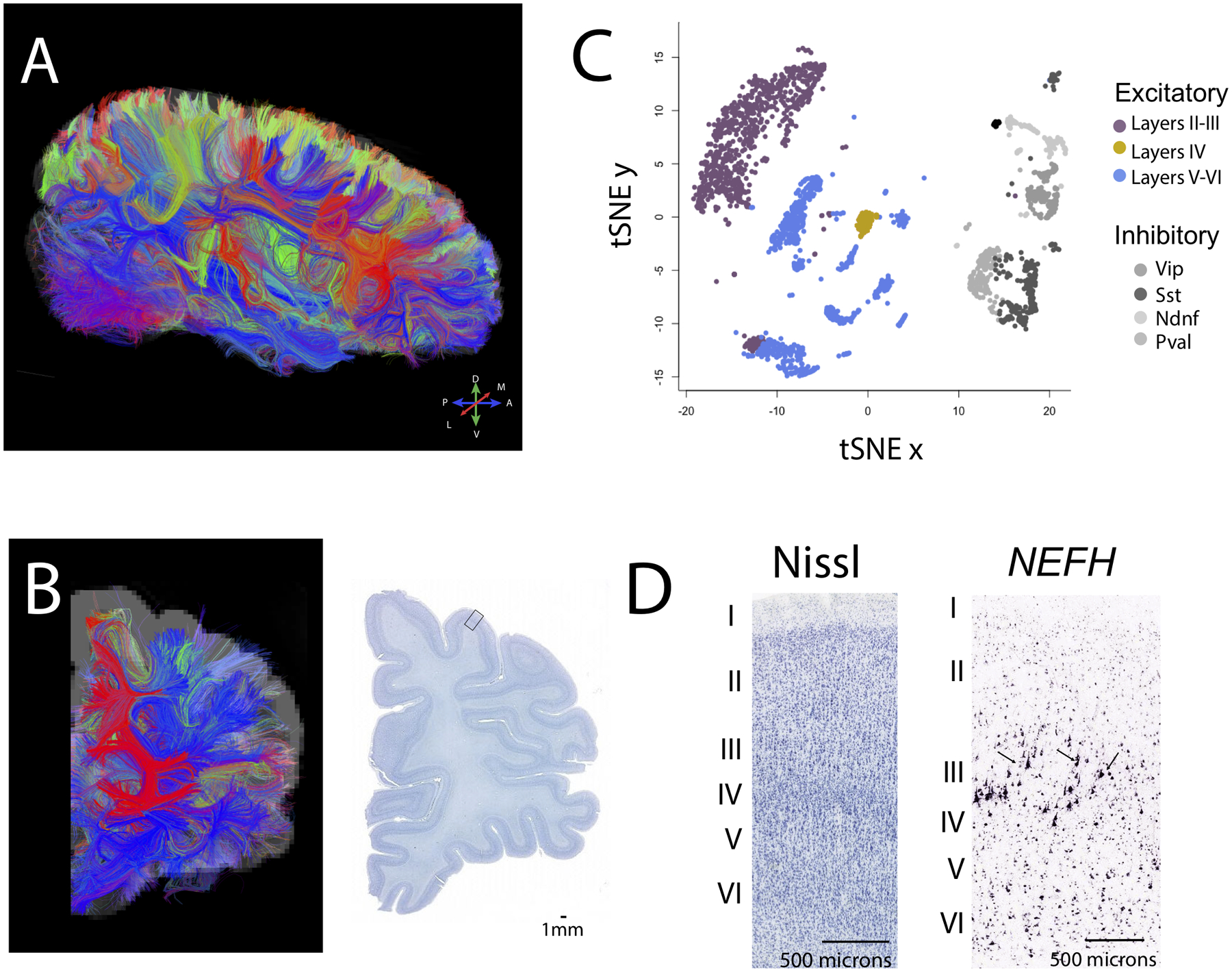
The integration of diffusion MR tractography with transcriptional and anatomical variation can be used to identify conservation and variation in connectivity patterns in humans relative to other species. Diffusion MR tractography viewed from sagittal (A) and (B) coronal slices show fibers (i.e., tracts) that course through the white matter. Color-coding of fibers represent their direction according to the map shown in A (e.g., fibers coursing across the (A) anterior to (P) posterior axis are in blue). (C) A tSNE plot of single cells from methylome sequencing highlights cell populations across layers of the frontal cortex of humans. These data can be used to compare relative populations of cell types across species. (D) Close-up view through the cortex identifies cell populations based on cytoarchitecture (Nissl-stained) and gene expression (NEFH). The expression of NEFH identifies large pyramidal neurons in layer III (arrows). Quantitative comparisons of diffusion MR tractography, single cell sequencing, and neuronal populations based on cytoarchitecture and gene expression can be used to identify conservation and variation in connections across species. Diffusion MR scans and in situ hybridization data are made available on the Allen Brain Institute for science. The diffusion MR scan at 900 μm resolution and Nissl-stained sections are from a 34 year old female brain (150). Single cell methylome sequencing data are from 107. The Nissl-stained section shown in D is taken from the box shown in B. Abbreviations: M: Medial; L: lateral; D: Dorsal; V: ventral.