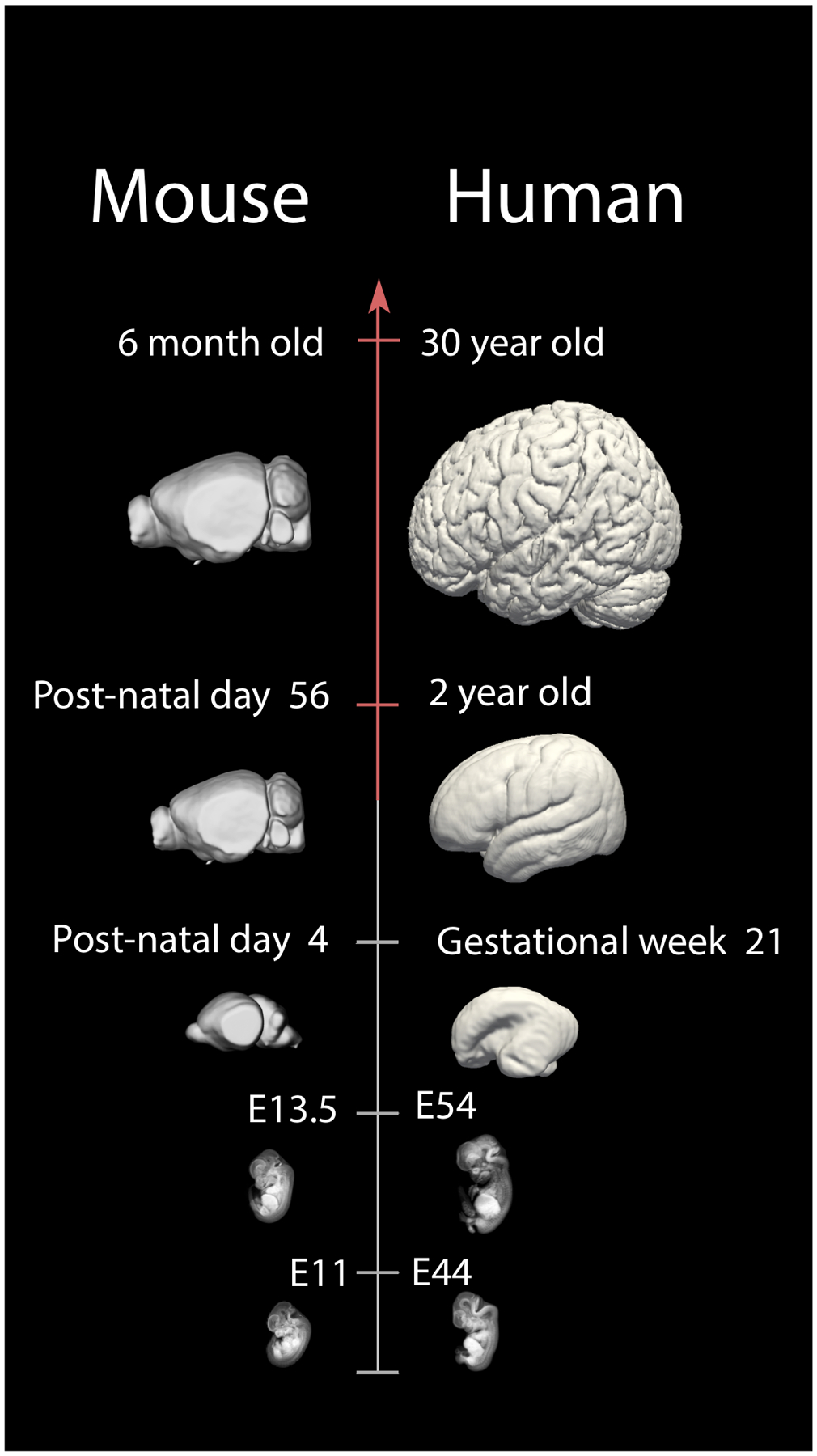
Figure 2.

A schematic illustrates examples of corresponding time points between humans and mice throughout fetal and postnatal ages. For example, a mouse at embryonic day (E) 11 is roughly equivalent to a human at E44. A post-natal day 4 mouse equates to a human fetus at gestational week 21, and a 6 month-old mouse is roughly equivalent to a human at 30 years of age. These data will enhance translational work from model systems to humans by finding corresponding ages throughout development and adulthood. Micro CT-scans of prenatal mice are from 151. Structural MR scans of humans at ED 44 and 54 are from the multi-dimensional human embryo project (http://embryo.soad.umich.edu.proxy.library.cornell.edu/index.html). Smooth surfaces were made from human MRI structural scans. Human and mice brains other than those from the Multi-dimensional project and 151 are made available from the Allen Brain Atlas.
