Figure 3.

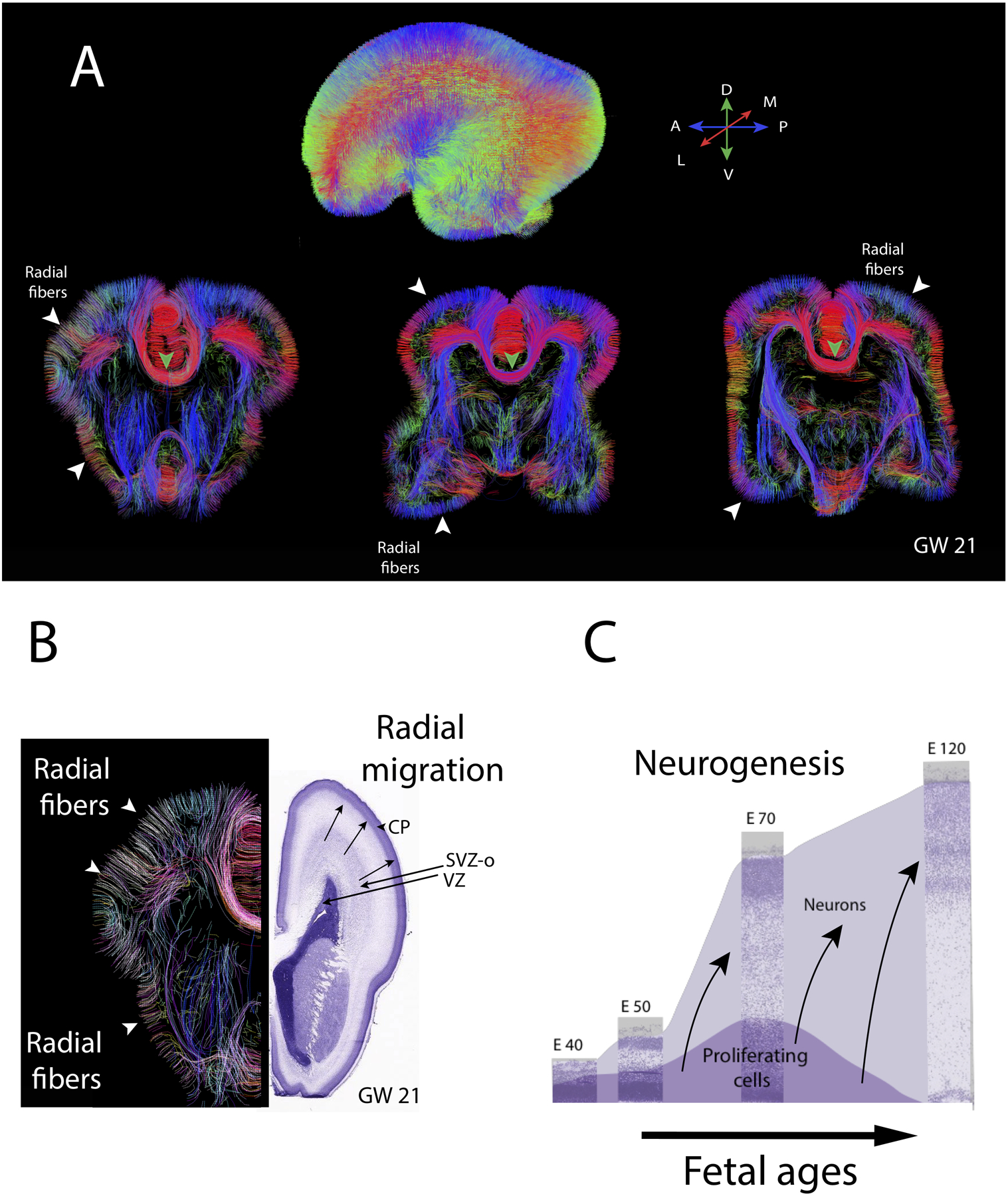
Diffusion MR tractography through the brain of a human fetus at gestational week (GW) 21. (A) Whole brain diffusion MR tractography highlight fibers coursing in different directions with the color-coding highlight the average direction of fibers (see map). Coronal slices through the brain highlight radially aligned fibers (i.e., putative radial glia) coursing from the proliferative zones to the outer surface of the developing cortex (white arrow-heads) as well as emerging pathways such as the corpus callosum (green arrow-heads). (B) Diffusion MR tractography from the left hemisphere show radial fibers coursing from the proliferative zone to the outer surface of the cortex. Nissl-stained sections through the right hemisphere show cell dense proliferative cells. These proliferative zones include the ventricular zone (VZ) and a subventricular zones (inner and outer; SVZO). Over the course of cortical neurogenesis, newly born neurons exiting the proliferative pools migrate radially along radial glia to the developing cortex called the cortical plate (CP). (C) Timetable of cortical neurogenesis at different ages represent the population of proliferative and neurons over the course of development. Neurons are produced over time and migrate radially to the outer surface of the cortex. Nissl-stained sections of sections of the developing cortex of macaques at different ages are in the background from embryonic (E) from E40 to E120. Over the course of fetal neurogenesis, the progenitor pool amplifies concomitant with newly born neurons cells exiting the proliferative zones to migrate to the outer surface of the cortex. These newly born neurons migrate to the outer surface of the cortex, and they are guided by radial glial cells as they migrate from the proliferative pool to the outer surface of the cortex. Neurons generated later migrate previously generated neurons (arrows). The diffusion MR scan of a 21GW fetus is from the Allen Institute for Brain Science. Abbreviations: A: Anterior; P: Posterior; M: Medial; L: lateral; D: Dorsal; V: ventral.
