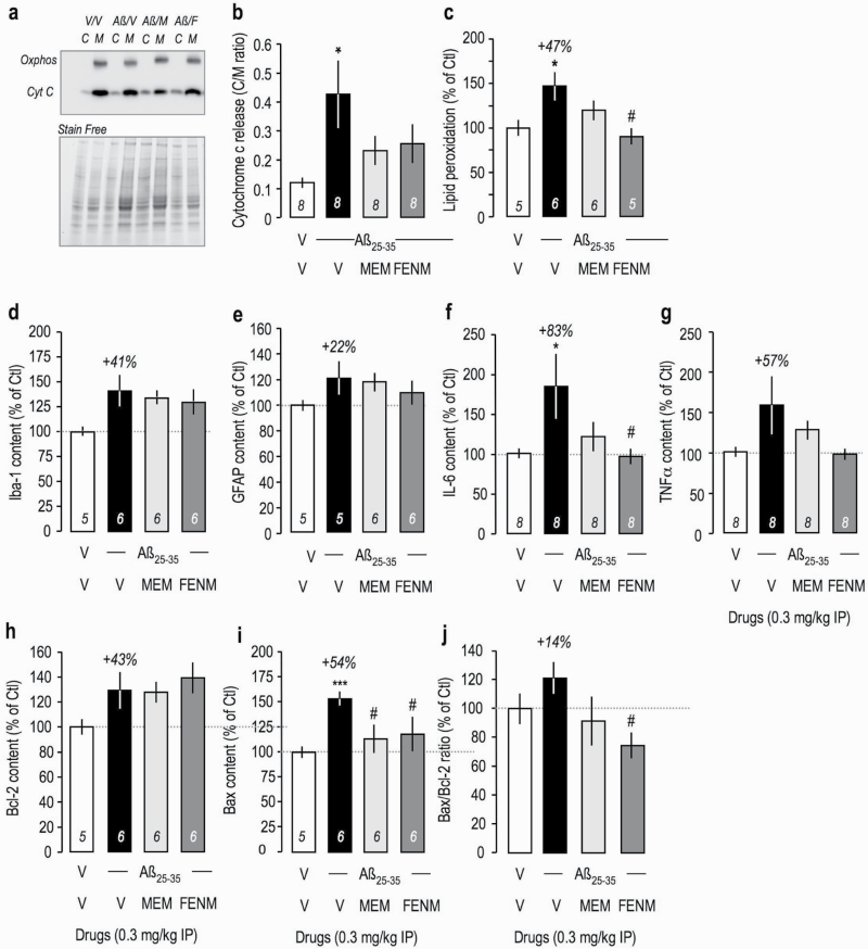
Figure 5.
Protective effects of Memantine and FENM administered at 0.3 mg/kg IP on Aβ 25-35-induced (a–c) oxidative stress and mitochondrial alteration and (d) Iba-1, (e) GFAP, (f) IL-6, (g) TNFα, (h) Bcl-2, and (i) Bax contents measured by ELISA in the mouse hippocampus. (a–b) Cytochrome c release from mitochondria to the cytosol in cortex extracts. (a) Typical blots showing oxphos-complex IV subunit I mitochondrial marker and cytochrome c labeling. Normalization was done with stain free total protein content in each band. (b) Quantification. (c) Measure of lipid peroxidation level in mouse cortex extracts. ELISA assays were done 16 days after ICV injection (d–e, h–i) or 5 days after ICV injection (f–g). (j) Bax/Bcl-2 ratio. ANOVA: F(3,31) = 3.05, P < .05 (b). F(3,21) = 4.33, P < .05; F(3,22) = 2.53, P > .05 (d); F(3,21) = 1.06, P > .05 (e); F(3,31) = 3.06, P < .05 (f); F(3,31) = 2.10, P > .05 (g); F(3,22) = 2.00, P > .05 (h); F(3,22) = 3.37, P < .05 (i); F(3,22) = 0.763, P > .05 (j). *P < .05, ***P < .001 vs (V+V)-treated group; #P < .05 vs (V+Aβ 25–35)-treated group; Dunnett’s test.

