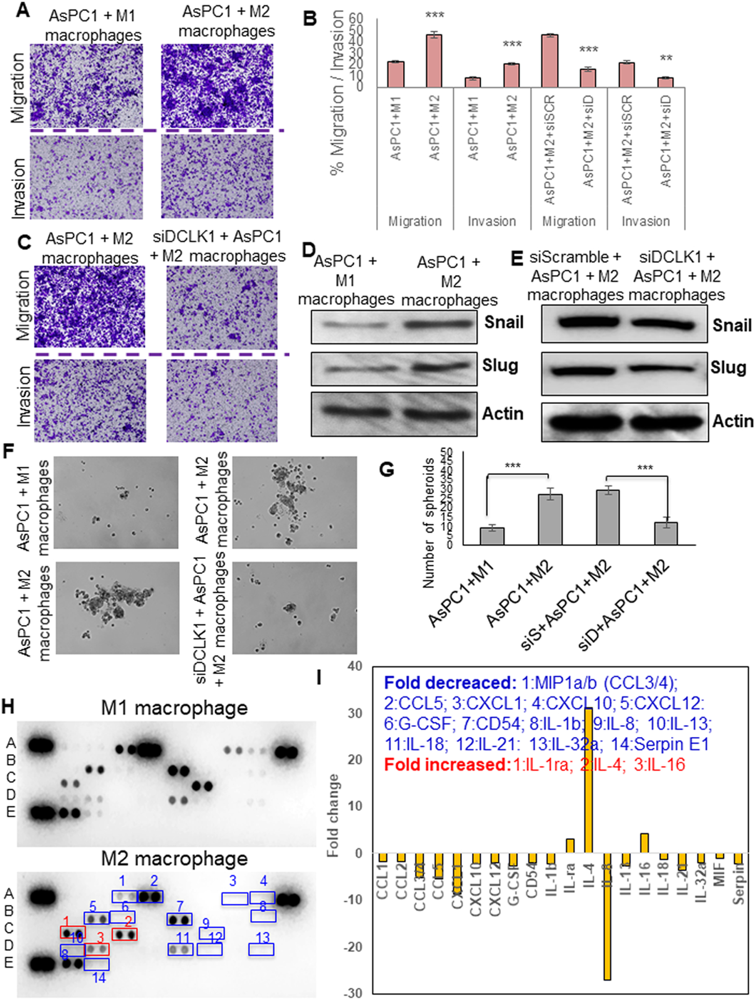
Fig. 4: DCLK1-isoform2 mediated M2-macrophages enhance PDAC aggression via soluble secretory factors.

A) Migration and invasion of AsPC1 cells from M1-macrophage or M2-macrophage dual-culture. B) Bar graph represents % tumor cell migrated or invaded. C) Migration and invasion of AsPC1 cells either treated with siScramble or siDCLK1 from M2-macrophage dual-culture. D) Protein expression of SNAIL and SLUG in the AsPC1 cells dual cultured with M1 or M2-macrophages. E) Protein expression of SNAIL and SLUG in the AsPC1 cells treated with siScramble or siDCLK1 from a dual culture experiment with M2-macrophages. F) Self-renewal of AsPC1 cells from M1-macrophage or M2-macrophage dual-culture; self-renewal of AsPC1 cells treated with siScramble or siDCLK1 from M2-macrophage dual-culture.G) Bar graph represents the number of spheroids formed. H) Human cytokine array by dot blot analysis, conditioned media collected from M1 or M2 macrophages utilized for the array. Cytokines/chemokines with 2-fold differences in their increase or decrease were numbered in the blot and named. I) Arrays were quantified using the densitometric analysis to represent a fold change in a bar graph. All quantitative data are expressed as means ± SD of a minimum of three independent experiments. P values of <0.05 = *, <0.01 = **, and 0.001 = *** were considered statistically significant.
