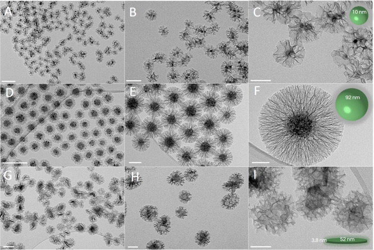
Figure 6.
Representative transmission electron microscopy micrographs with increasing magnification of three different types of core–shell magnetic nanoparticles with radially aligned silica pores: superparamagnetic iron oxide nanocrystals (A–C), superparamagnetic nanocrystal clusters (D–F), and hard-magnetic barium hexaferrite nanoplatelets (G–I), generated after the coating process by using the general procedure. Scale bars: 200 nm (A, D, G); 100 nm (B, E, H); 50 nm (C, F, I).

