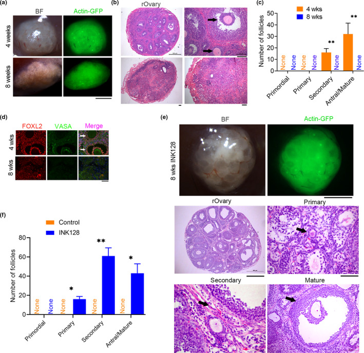
FIGURE 1.

INK128 maintains follicular development in the reconstituted ovaries (rOvary) of young recipient C57BL/6 mice. (a) Morphology of the rOvary four or 8 weeks following transplantation of PGCs aggregated with E12.5 gonadal somatic cells into young recipient C57BL/6 mice (2–3 months old, an average of 10 weeks old) treated with or without INK128 (control). BF, microscopic image under bright field; actin‐GFP indicates donor cell sources from mice carrying actin‐GFP. Scale bar = 1 mm. (b) Histology of rOvary sections by H&E staining indicating various follicles 4 weeks, but the absence of follicles 8 weeks following transplantation. The black arrows refer to secondary and mature follicles. Scale bar = 100 μm. (c) Number of follicles at various developmental stages in the rOvary four or 8 weeks after transplantation of PGCs. n = 3. (d) Immunofluorescence of FOXL2 indicating granulosa cells in the follicles and VASA to label oocytes. Scale bar = 50 μm. (e) Follicles maintained in the rOvary of recipient mice by treatment with INK128 for 8 weeks. Upper panel, morphology of rOvary; scale bar = 1 mm. Bottom panel, histology of rOvary sections by H&E staining displaying various follicles. Scale bar = 500 μm. Shown also are primary, secondary, and mature follicles indicated by black arrows at higher magnifications (scale bar = 100 μm). (f) Number of follicles at various developmental stages in the rOvary 8 weeks after transplantation of PGCs in the recipient mice treated with INK128, compared with control recipient mice without INK128 treatment. Mean ± SEM. *p < 0.05, **p < 0.01
