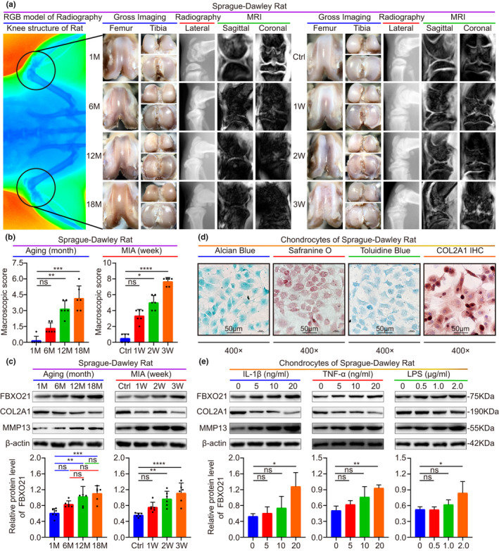
FIGURE 2.

FBXO21 accumulates in rat articular cartilage and chondrocytes with OA. (a) Gross imaging, plain radiograph, and magnetic resonance imaging (MRI) scans from aging (middle) and monosodium iodoacetate (MIA) (right) OA rat models. Radiography presents the whole knee structure (left). M: Month; Ctrl: Saline Controls; W: Week. (b) Macroscopic score. Immunoblotting (upper) results of FBXO21, COL2A1, and MMP13 and the quantification (lower) of FBXO21, with β‐actin as the endogenous control, in the (c) aging and MIA OA rat models and in (e) chondrocytes stimulated with interleukin (IL)‐1β, tumor necrosis factor (TNF)‐α, and lipopolysaccharide (LPS). (d) Identification of rat chondrocytes using Alcian Blue, Safranin O, or Toluidine Blue and immunohistochemistry (IHC) staining of COL2A1. Data are presented as the mean ± SD; ns: not significant, *p < 0.05, **p < 0.01, ***p < 0.001, ****p < 0.0001
