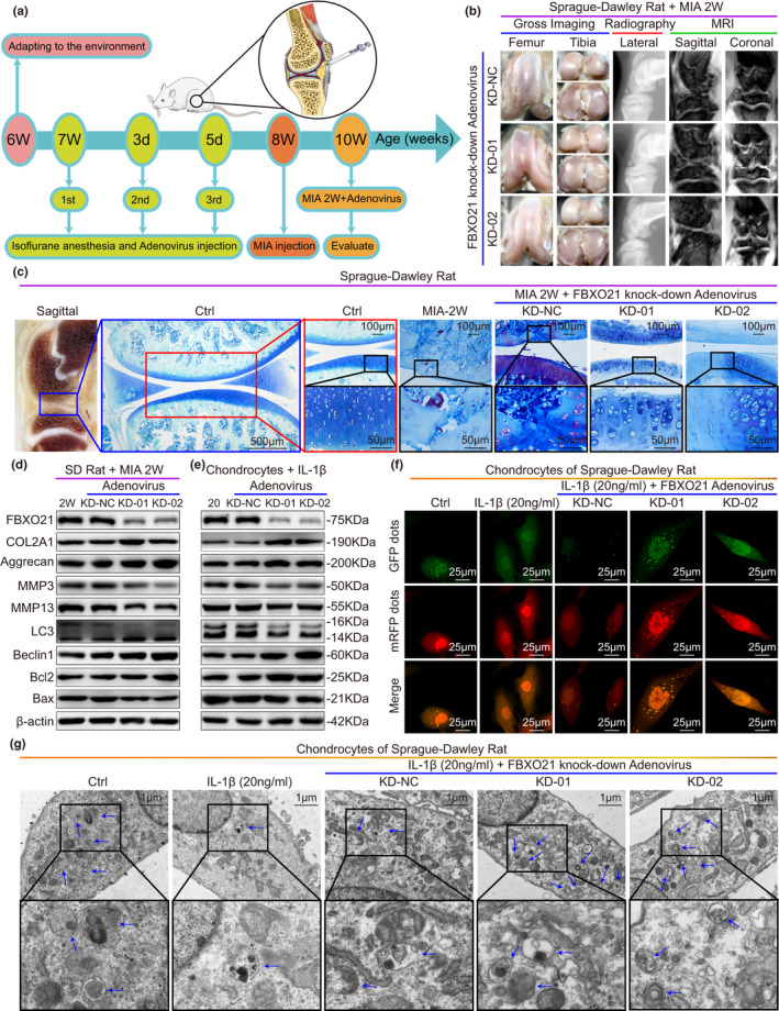
FIGURE 3.

FBXO21 knockdown suppresses OA‐related degeneration in MIA‐treated rats and IL‐1β‐treated rat chondrocytes. (a) Experimental diagram of the monosodium iodoacetate (MIA) OA rat models treated with adenovirus (Ad) carrying FBXO21‐specific knockdown (KD) shRNA and overexpression (OE) plasmid. Rats were evaluated at age of 10 weeks. 1st: first; 2nd: second; 3rd: third; d: day. (b) Gross imaging, plain radiograph, and MRI scans of MIA‐2W OA rats. KD‐Ad‐shRNA‐NC (KD‐NC), KD‐Ad‐shRNA‐FBXO21‐01 (KD‐01), and KD‐Ad‐shRNA‐FBXO21‐02 (KD‐02) groups were formed. NC: negative control. (c) Toluidine Blue staining of rat knee joints and their sagittal section (left). Boxed regions represent higher magnification. Ctrl: Saline Controls. Immunoblotting results of FBXO21, COL2A1, Aggrecan, MMP3, MMP13, LC3, Beclin1, Bcl2, and Bax, with β‐actin as endogenous control, in (d) rat knee cartilage and (e) rat chondrocytes. 2 W: MIA‐2 W; 20: 20 ng/ml of interleukin (IL)‐1β. (f) mRFP‐GFP‐LC3 adenovirus double label in chondrocytes after knockdown of FBXO21. mRFP (red) indicates autolysosomes (ALs); Merge (yellow) indicates autophagosomes (APs). (g) Ultrastructural features of APs in the chondrocytes observed using transmission electron microscopy. Black box represents higher magnification. Blue arrows indicate APs
