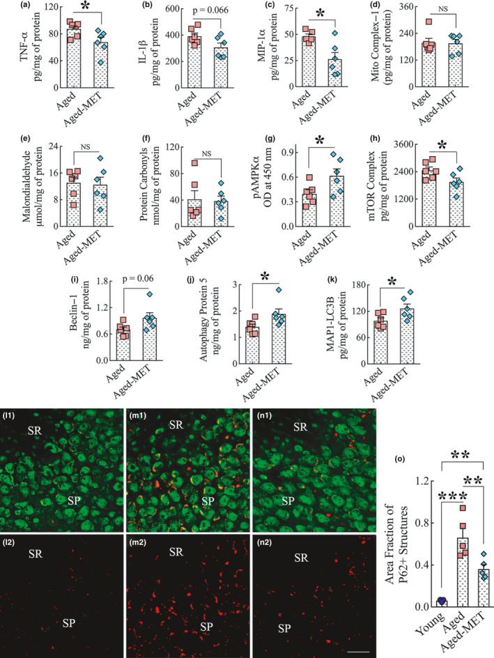
Figure 6.

Two weeks of MET treatment to 18‐month‐old mice reduced proinflammatory markers, activated AMPK, inhibited the mammalian target of rapamycin (mTOR), and enhanced autophagy in the hippocampus without altering the concentration of mitochondrial complex 1, or the oxidative stress markers malondialdehyde and protein carbonyls. The bar charts a‐c show a reduced concentration of proinflammatory markers TNF‐α (a), IL‐1β (b), and MIP‐1α (c) in the hippocampus of the MET‐treated aged mice compared to untreated aged mice. The bar charts (d–f) show that the concentration of mitochondrial complex I (d), malondialdehyde (e), and protein carbonyls (f) in the hippocampus did not differ between untreated aged mice and MET‐treated aged mice. The bar charts in g–h show that MET treatment increased the concentration of pAMPKα (g) and decreased the level of mTOR complex (h) in the hippocampus. The bar charts i‐k show MET treatment enhanced the levels of autophagy‐related proteins beclin‐1 (i), autophagy protein 5 (j), and microtubule‐associated protein 1 light chain 3 beta (MAP1‐LC3B; k) in the hippocampus. *p < 0.05. l1‐n2 shows representative images of p62 and NeuN dual immunofluorescence in the CA3 subfield of the young mouse (l1–l2), untreated aged mouse (m1–m2), and the aged mouse that received 10 weeks of MET treatment commencing in late middle age (n1, n2) mice. The bar chart (o) compares the area fraction of p62+ structures in the CA3 subfield of the hippocampus across three groups. Note that the area fraction of p62+ structures is decreased in MET‐treated aged mice compared to the untreated aged mice. SP, stratum pyramidale; SR, stratum radiatum; NS, not significant. **p < 0.01; ***p < 0.001. Scale bar, l1‐n2 = 25 μm
