FIGURE 4.

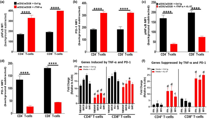
Recombinant IL‐37 treatment opposes TNF‐α signaling in aged T‐cells. Naïve CD4+ and CD8+ T‐cells were purified from aged (24 months old) C57BL/6 mice via MACs selection and stimulated in vitro with αCD3/αCD28 in the presence of Control Ig, rTNF‐α, or rTNF‐α + rIL‐37. (a, c) After 10’ of stimulation, phospho‐flow cytometry was performed to determine NF‐κB activation. (b, d) After 3 days of stimulation, the surface expression of PD‐1 on aged T‐cells was determined using flow cytometric analysis. (e, f) Naïve T‐cells were purified as described above and stimulated with Control Ig or rIL‐37 for 4 h. After the stimulation period, qPCR analysis was performed to ascertain the expression levels of genes involved in T‐cell activation (IFN‐γ, TBK1, IRF3) and inhibition (TMEM16F, GM130, SHP2, and PD1). Importantly, the genes chosen for assessment are regulated by TNF‐α and PD‐1 signaling. Significance was determined using a Student's t test relative to αCD3/αCD28 + Control Ig (a–d) and media +Control Ig (e, f) treated groups. For a–d, means ± SD are shown with ****p < 0.0001. For (e) and (f), a=*p < 0.05, b=**p < 0.01, c=***p < 0.001, and d=****p < 0.0001 where gene expression levels observed in Control Ig‐treated aged T‐cells were used as the positive control for each gene tested. n = 9 mice/group with 3 independent experiments conducted
