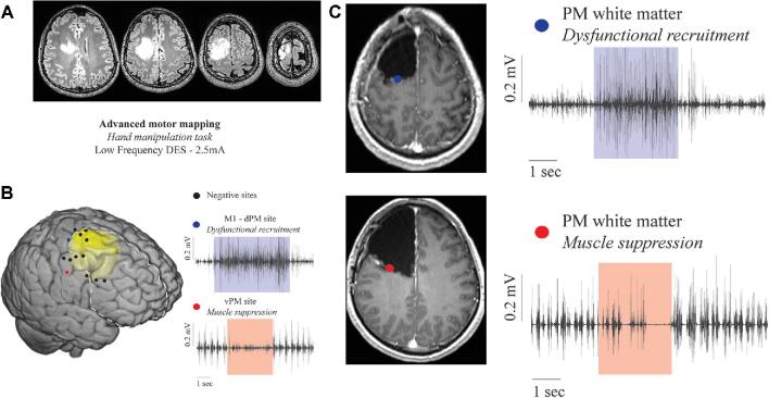
FIGURE 3.
Representative case of a 39-yr-old male with a previous history of focal seizures presented with a low-grade tumor involving the right prefrontal area. The patient was operated under asleep-awake-asleep anesthesia. The hMT was performed on the cortical and subcortical levels to assess the praxis network. A, Preoperative axial FLAIR pictures. B, MNI template in which the tumor volume was reported in yellow. Cortical sites tested during hMT with LF (3 mA, bipolar probe) are reported. Sites where no interferences were evoked are represented with black dots. The only site where dysfunctional recruitment was induced is reported as a blue dot (the EMG trace recorded from APB shows modification of the pattern of muscle activation during stimulation). The only site where a muscle suppression was induced is reported as a red dot (the EMG trace recorded from APB shows a significant reduction in motor unit recruitment). The light blue and red boxes highlight the stimulus application. C, Subcortical stimulation with LF during hMT identifies the posterior subcortical boundaries of resection. The upper panel shows a site where dysfunctional recruitment was induced (blue dot superimposed on an axial T1-post gadolinium postoperative MR) (EMG trace of APB showing modification of muscle pattern of activation). The lower panel shows a site where muscle suppression was induced (red dot superimposed on an axial T1-post gadolinium postoperative MR) (EMG trace of APB showing an almost complete suppression of muscle activation).

