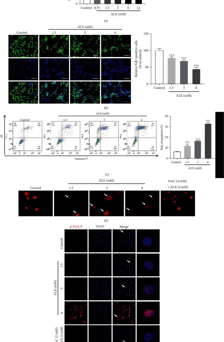
Figure 4.

ALX significantly induced injury toward pancreatic β-cells. (a) The cytotoxicity of ALX (0.75 to 12 mM, 24 h) toward MIN6 cells was evaluated by using an MTT assay. (b) EdU incorporation was evaluated after treatment of cells with vehicle and ALX (1.5, 3, and 6 mM) for 24 h. The EdU-positive cells in each group were quantified as the percentage of those in the control group. (c) The impact of vehicle and ALX (1.5, 3, and 6 mM, 24 h) on MIN6 cell apoptosis was detected by Annexin V-FITC/propidium iodide (PI) staining. (d) Representative comet tail images showing the DNA damage response after treatment with vehicle and ALX (1.5, 3, and 6 mM) for 24 h. (e) Representative confocal images (scale bar: 50 μm) of double-stained cells subjected to the same treatments and stained for p-H2A.X (red) and with DAPI (blue). The data represent the mean ± SD (n = 3). ∗∗p < 0.01 and ∗∗∗p < 0.001 compared with the control group.
