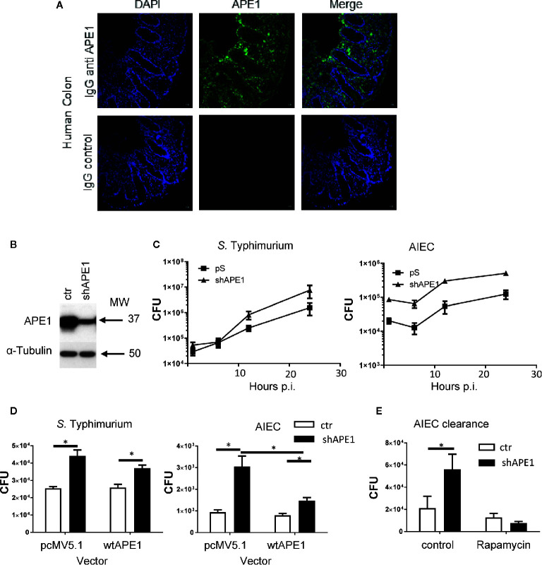
Figure 1.
Numbers of intracellular bacteria in intestinal epithelial cells are negatively regulated by APE1. (A) To confirm that APE1 was expressed by human epithelial cells, human colonic biopsy specimens were stained with DAPI or goat anti-APE1 followed by an anti-goat secondary antibody and imaged. Staining can be detected in both the epithelium and lamina propria. (B) T84 cells containing short hairpins to APE1 have reduced levels of APE1 protein compared to cells containing a control vector. (C) T84 cells with normal or suppressed levels of APE1 were infected with S. Typhimurium at MOI 10 or AIEC at MOI 100 for 1 h and assayed for intracellular bacteria using the gentamicin inhibition assay. N= 6-8 pooled from two independent experiments. (D) To ensure APE1 specificity of our findings in cells containing shAPE (targeting intron), APE1 levels were complemented by transfecting HT-29 cells containing shAPE with a vector expressing wild type (wt) APE1 or a control vector (pcMV5.1) and then infected with S. Typhimurium (MOI 10) or AIEC (MOI 100) for 1 h in two independent experiments with each 3-5 replicates. Control (ctr) cells with normal levels of APE1 had lower levels of intracellular bacteria with no further reduction by the transfection to increase wild type APE1 (open bars). Inhibition of APE1 by shAPE1 (black bars) increased intracellular bacteria which decreased after complementing with wild type APE1, particularly for AIEC. (E) To exclude the possibility that increased intracellular bacteria were caused by impaired autophagy, T84 cells were pretreated with rapamycin before assaying for bacterial internalization (expressed as CFU) to show that APE1-deficient cells have functional autophagy. All error bars are represented as SEM. *p < 0.05

