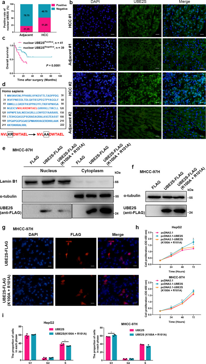
Fig. 2.
UBE2S enters the nucleus through nuclear localization signals and promotes HCC proliferation. a The positive rate of nuclear UBE2S in human HCC and adjacent tissues evaluated by immunohistochemistry. (n = 80 pairs). b Immunofluorescence detection of UBE2S expression in human HCC and adjacent tissues. Scale bars: 100 μm. c OS rates of HCC patients with positive or negative nuclear UBE2S content were evaluated by Kaplan–Meier analysis (n = 80 cases). d Bioinformatics analysis of the NLS in UBE2S and a strategy for its mutational analysis. e–f UBE2S-FLAG expression in the nuclear fractions of cell lysates and total lysates in MHCC-97H cells transiently transfected with UBE2S-FLAG NLS mutation plasmids assessed by western blotting. g Distribution of UBE2S-FLAG in MHCC-97H cells following mutation of the NLS detected by immunofluorescence analysis. h–i Effects of NLS mutations in UBE2S on cell proliferation and cell cycle progression determined by CCK-8 and flow cytometry assays. Two-tailed Student’s t tests were used to test the significance of differences between two groups; data are represented as mean ± SEM (h–i). Kaplan–Meier curves of overall survival of HCC patients were determined by the log-rank test (c). *P < 0.05, **P < 0.01

