Figure 1.

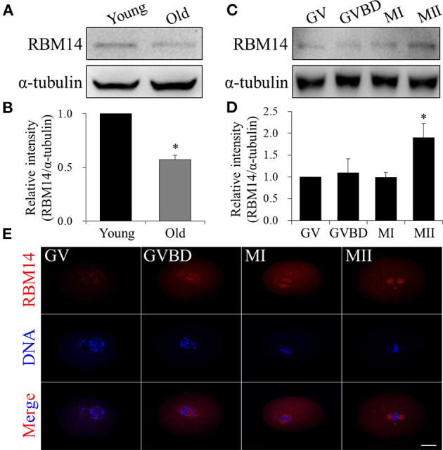
RBM14 expression in oocytes from aged mice and the expression and localization patterns of RBM14 during mouse oocyte maturation. (A) Western blot analysis showing RBM14 protein levels in oocytes from young (6–8 weeks) and old (42–45 weeks) mice. (B) Normalized band intensities: RBM14/α-tubulin. RBM14 protein levels were significantly lower in oocytes obtained from old mice compared to young mice. (C) RBM14 protein levels at GV (0 h), GVBD (2 h), MI (8 h), and MII (12 h) stages of young mice were detected by Western blot analysis; α-tubulin was the loading control. (D) Normalized band intensities: RBM14/α-tubulin. RBM14 protein levels were significantly higher in oocytes at MII stage compared to GV stage. *P < 0.05. (E) Immunofluorescence staining: red = RBM14 antibody, blue = DNA (Hoechst 33342). Scale bar = 20 μm. Data are mean ± SEM of three independent experiments. *P < 0.05.
