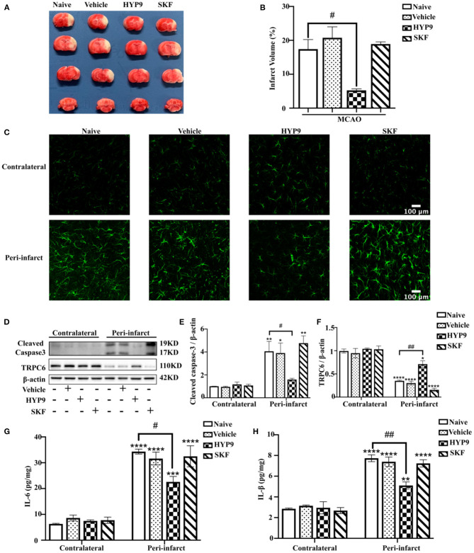
Figure 3.
Reducing IR-mediated TRPC6 downregulation with HYP9 alleviates ischemic damage in mice. Vehicle (DMSO), 5 μg HYP9, or 20 μg SKF were injected into left ventricles of mice 24 h before sham or MCAO operation. (A) TTC-stained cerebral slices indicated the infarct size in mice. (B) Quantification of infarct volumes after ischemia. Data were shown as mean ± SEM (n = 3). #p < 0.05 vs. MCAO + Naive group. (C) Representative images of astrocytes population with GFAP (green) staining in contralateral or peri-infarct cortices in MCAO mice (Scar bar: 100 μm; 400× magnification). (D) Western blot detecting TRPC6 and cleaved caspase-3 in mouse cortices. β-actin was used as a loading control. (E,F) Quantifications of cleaved caspase-3 and TRPC6 protein levels shown in (D). Data were shown as mean ± SEM (n = 3). (G,H) The release of IL-6 and IL-1β was determined by ELISA assay. Data were shown as mean ± SEM (n = 3). *p < 0.05, **p < 0.01, ***p < 0.001, ****p < 0.0001 vs. MCAO + Naïve group in contralateral cortices; #p < 0.05, ##p < 0.01 vs. MCAO + Naive group in peri-infarct area.

