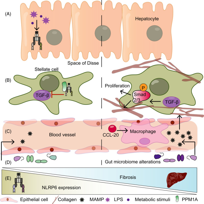
FIGURE 3.

NLRP6 in the liver. (A) Stimulation of hepatocytes with LPS or palmitic acid is accompanied by increased Nlrp6 expression. (B) In hepatic stellate cells, NLRP6 interacts with PPM1A to inhibit transforming growth factor‐β (TGF‐β). This, in turn, leads to reduced phosphorylation of Smad2/3 and reduced proliferation and collagen expression. (C) NLRP6 prevents the infiltration of macrophages into the liver by reducing C‐C chemokine ligand 20 (CCL20) levels. (D) Whole‐body Nlrp6 deletion leads to a dysregulated microbiota, which increases the influx of TLR ligands in the circulation, thereby promoting fibrosis. (E) In fibrotic livers, Nlrp6 expression is decreased, the functional implications of which remain unknown to date
