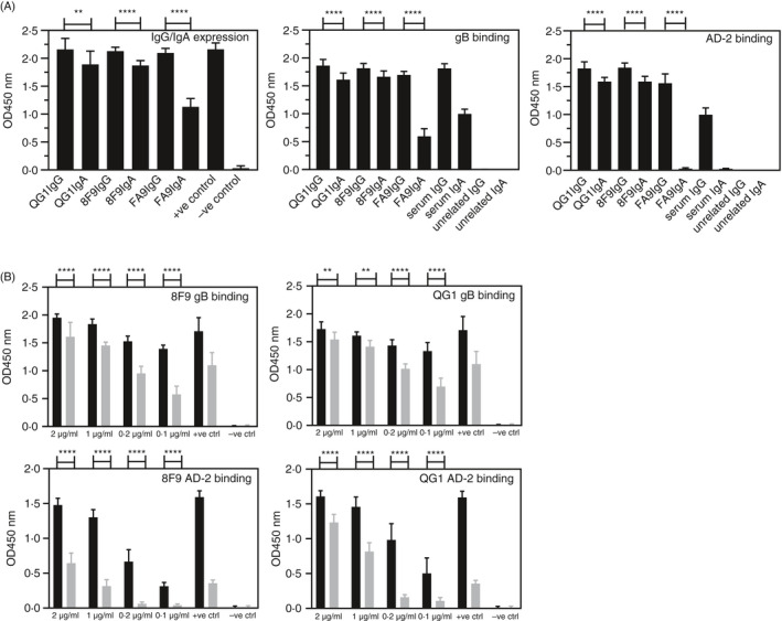
Figure 6.

Recombinant antibodies expressed as human IgG and IgA bind gB and AD‐2 epitope of HCMV. (A) Expression of monoclonal recombinant antibodies QG1 IgG1, QG1 IgA1, 8F9 IgG1, 8F9 IgA1, FA9 IgG1 and FA9 IgA1 by transient transfection and ELISA of undiluted culture supernatants and binding to gB and AD‐2. +ve control, diluted human serum; –ve control, undiluted culture supernatant from untransfected cells; unrelated IgG and IgA are control human mAbs. (B) Binding and titration of concentration‐matched mAbs QG1 IgG1 (black bars), QG1 IgA1 (grey bars), 8F9 IgG1 (black bars), 8F9 IgA1 (grey bars) to gB and AD‐2 epitope of HCMV represented as OD values with human serum as positive control and unrelated IgG and unrelated IgA as negative control. Statistical differences between the mean OD values of IgG and IgA expression and for binding to gB and AD‐2 epitope were obtained from the Mann–Whitney test (**P < 0.01; ****P < 0.0001)
