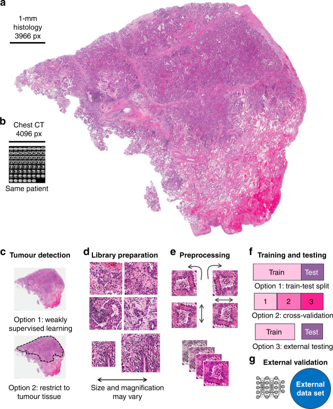
Fig. 1. Consensus pipeline of deep learning in pathology.
a Routine histology image of lung cancer (from The Cancer Genome Atlas (TCGA) and The Cancer Imaging Archive (TCIA)). b Size comparison (in terms of pixels) of a chest CT scan of the same patient. c Consensus image-processing pipeline. First, either the whole slide or just the tumour region is tessellated into smaller image tiles. d These tiles comprise an image library, similar to the library preparation (prep.) in genome sequencing. e Tiles are preprocessed to achieve rotational constancy and augment the dataset. f Deep- learning classifiers are developed and deployed by splitting the patient cohort into a training and testing set, by using cross-validation or by having multiple cohorts available for training and testing. g Ideally, an additional external dataset is used for validation of the resulting classifier.

