Figure 2.

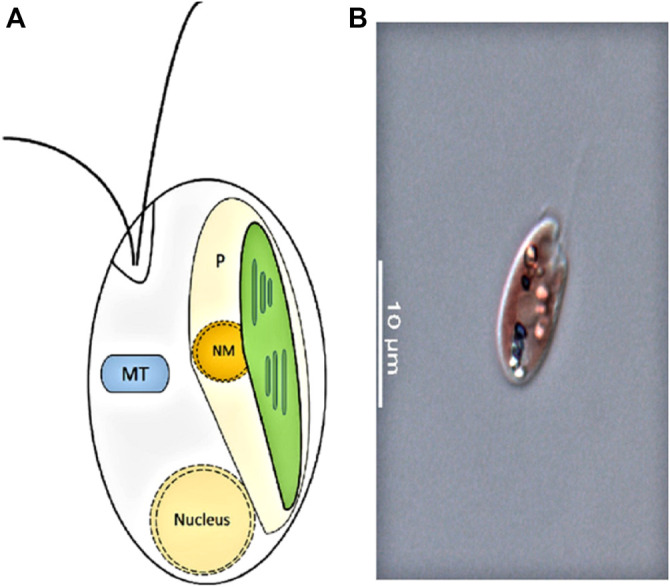
(A): Cryptophyte cell structure. P, plastid; NM, nucleomorph; MT, mitochondrion (adapted from Hoef-Emden 2008). (B): Photo of a cryptophyte Rhinomonas nottbecki n. sp. taken by Janne-Markus Rintala.

(A): Cryptophyte cell structure. P, plastid; NM, nucleomorph; MT, mitochondrion (adapted from Hoef-Emden 2008). (B): Photo of a cryptophyte Rhinomonas nottbecki n. sp. taken by Janne-Markus Rintala.