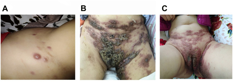
Figure 2.
The evolution of skin metastases.
Notes: In September 2019, the skin of the hypogastrium and perineum was red and swollen with a rubbery appearance, a rash-like swelling on the surface, and local fusion (A); In December 2019, the skin nodules had increased in size and involved the skin of the hypogastrium, left thigh, bilateral groin, and perineum. These nodes mixed together, and formed tiny open sores, or ulcers, on the surface of the nodes (B); After palliative treatment with FOLFIRI plus cetuximab and vemurafenib, the cutaneous nodules decreased in size (C).

