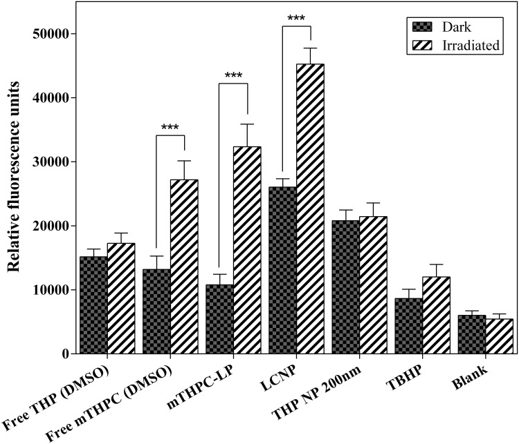
Figure 6.
Production of ROS in response to mTHPC loaded liposomes, free mTHPC (DMSO), THP loaded PLGA nanoparticles (200 nm), free THP (DMSO), and lipid-coated polymeric nanoparticles (LCNP). H2DCFDA (25 µM) was used as a free radical quenching fluorescent dye. The cells were incubated with the formulations or free drugs (at a corresponding mTHPC and THP concentration of 5 µM and 50 µM, respectively) for 4 h at 37°C. Subsequent radiation was performed at a light dose of 0.5 J.cm−2 (λ = 652 nm). Blank represents the non-illuminated cells whereas TBHP (50 μM) was used as a positive control. All the measurements were performed in triplicate and values were expressed as mean ± S.D (n=3). P values (p < 0.05) were considered significant and expressed as ***p< 0.001.

