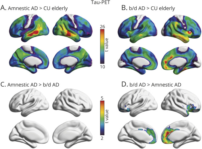
Figure 3. Topographic Distribution of Amyloid-β in Amnestic and Behavioral/Dysexecutive (b/d) Variants of Alzheimer Disease (AD).
(A) Strongest associations between [18F]MK6240 standardized uptake value ratio (SUVR) and amnestic AD were observed in lateral temporal, inferior parietal, precuneus, and posterior cingulate cortices. (B) Strongest associations between [18F]MK6240 SUVR and behavioral/dysexecutive AD were observed in the anterior cingulate, lateral temporal, frontal insula, and orbitofrontal cortices. (C) t Maps displaying significant differences between patients with behavioral/dysexecutive AD and patients with amnestic AD; after multiple comparisons correction with random field theory (p < 0.001), no results remained significant. (D) t Maps displaying significant differences between patients with behavioral/dysexecutive AD and patients with amnestic AD. Behavioral/dysexecutive subjects had greater tau-PET uptake in medial prefrontal, anterior cingulate, and frontal insular cortices. t Statistical parametric maps were corrected for multiple comparisons using a random field theory cluster threshold of p < 0.001. Age and sex were employed as covariates in each model. CU = cognitively unimpaired.

