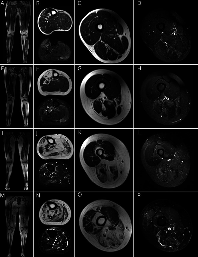
Figure 1. MRI Findings in GNE Myopathy.
Representative T1-weighted (T1W) coronal and axial MRI of lower legs and thighs and corresponding axial short tau inversion recovery (STIR) images in patients with GNE myopathy. (A–D) MRI of a patient early in the disease progression (lower extremity [LE] strength: 94%) shows (A) minimal changes on T1W images and (B) STIR hyperintensity of the anterior tibialis, extensor digitorum longus (EDL), extensor hallucis longus (EHL), and gastrocnemius medialis muscles, and minimal changes in the thigh muscles (C and D). (E–H) A patient with intermediate disease progression (LE strength: 73%) showing 4 stages of muscle involvement identified by MRI: stage I: muscles with no visible abnormalities on T1W or STIR soleus (F) and quadriceps (G) muscles; stage II: STIR hyperintense muscle with minimal or no fat infiltration: gastrocnemius medialis (F) and muscles of the medial and posterior compartments of the thigh (G and H); stage III: muscles with fat infiltration and STIR hyperintensity: tibialis posterior and peroneus brevis (F); stage IV: complete fat replacement: anterior tibialis, EHL, and EDL muscles (F). (I–L) Patient with more advanced disease progression (LE strength: 39%); MRI shows the majority of lower leg muscles at stage IV (J), and involvement of the medial and posterior thigh muscles (K, L). In the anterior thigh compartment, there is STIR hyperintensity of the rectus femoris and vastus intermedius (stage II), with no visible abnormalities of the vastus lateralis (L). (M–P) Patient with advanced disease progression (LE strength: 16%); most of the lower extremity muscles were at stage IV (M, N, O), except for portions of the vastus lateralis and medialis, which show STIR hyperintensity (O, P).

