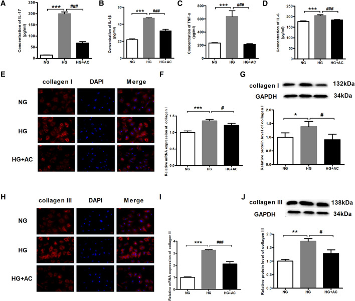
FIGURE 4.
Anthocyanin reduces myocardial fibrosis and the levels of inflammatory cytokines in cardiac fibroblasts from neonatal mice induced by high glucose. (A) Concentration of interleukin (IL)-17 detected by enzyme-linked immunosorbent assay (ELISA). (B) Concentration of IL-1β detected by ELISA. (C) Concentration of TNF-α detected by ELISA. (D) Concentration of IL-6 detected by ELISA. (E) The immunofluorescence staining of collagen I. Scale bar: 20 μm. (F) Relative expression of collagen I mRNA detected using quantitative reverse transcription PCR (qRT-PCR). (G) Relative protein level of collagen I. (H) The immunofluorescence staining of collagen III. Scale bar: 20 μm. (I) Relative expression of collagen III mRNA. (J) Relative protein level of collagen III. GAPDH served as an internal control. *p < 0.05 vs. NG, **p < 0.01 vs. NG, ***p < 0.001 vs. NG, #p < 0.05 vs. HG, ###p < 0.001 vs. HG. NG, normal glucose (5.5 mM); HG, high glucose (25 mM); HG + AC, high glucose (25 mM) + anthocyanin (250 μg/ml); n = 3.

