Fig. 1.

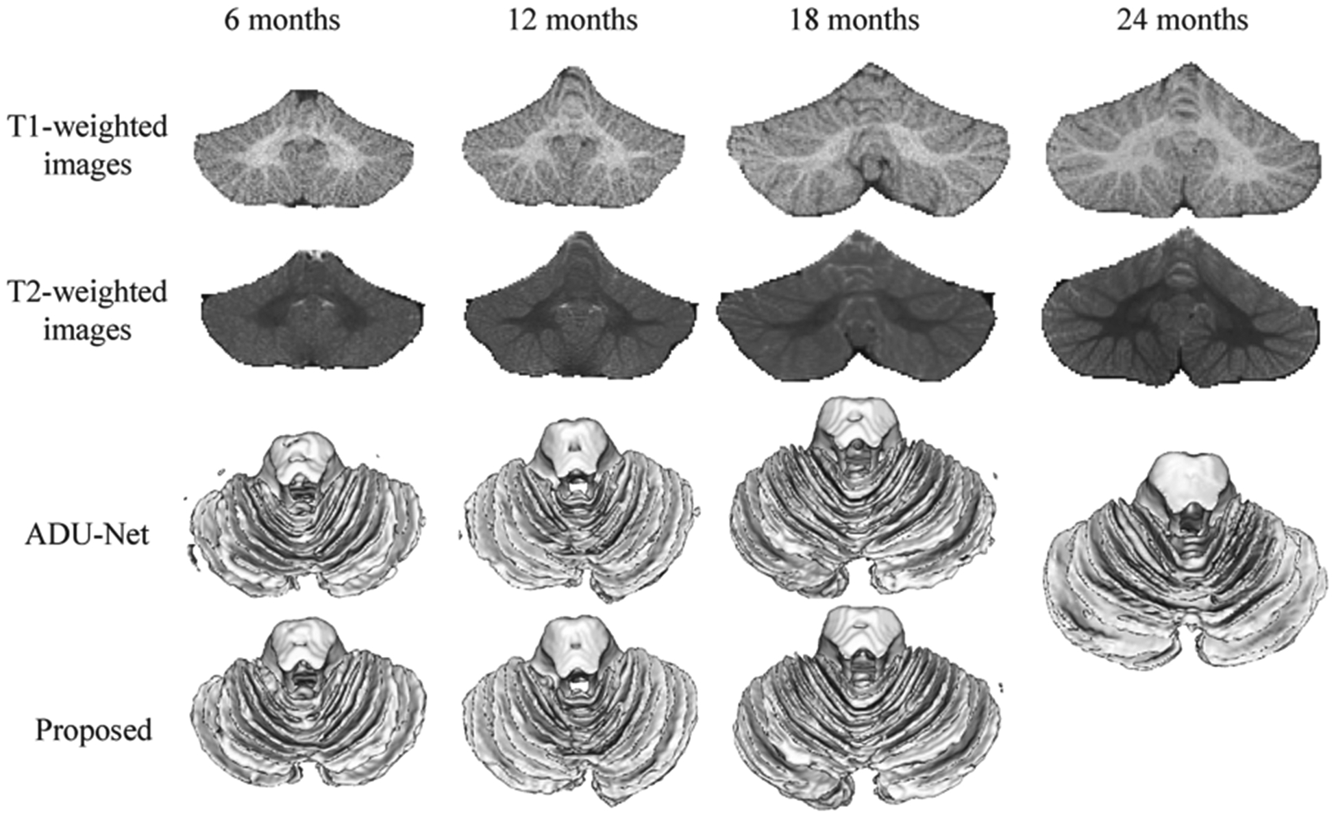
T1- and T2-weighted MRIs of the cerebellum at 6, 12, 18 and 24 months of age, with the corresponding segmentation results obtained by ADU-Net [9] and the proposed semi-supervised method.

T1- and T2-weighted MRIs of the cerebellum at 6, 12, 18 and 24 months of age, with the corresponding segmentation results obtained by ADU-Net [9] and the proposed semi-supervised method.